
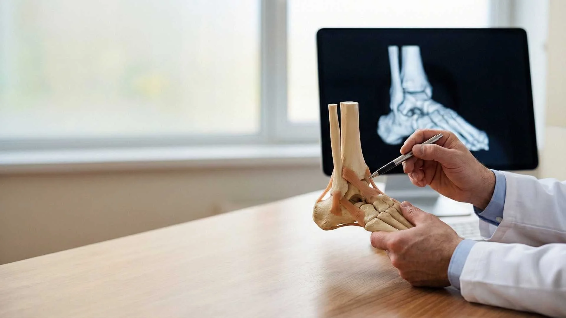
Ankle arthroscopy is a minimally invasive surgical technique used to diagnose and treat joint problems in the ankle (tibiotalar joint). Using small keyhole incisions (typically on the front or back of the ankle), orthopedic surgeons can insert a small camera and instruments to address joint issues. This technique avoids large incisions in an area of the body with thin skin and limited soft tissue coverage, reducing wound healing risks and expediting recovery.
Dr. Francisco Hernandez provides specialized ankle evaluations and arthroscopic surgeries at the modern medical facilities of Hospital Punta Mita, serving patients in Puerto Vallarta and the Riviera Nayarit.
When to Consider Ankle Arthroscopy? (Symptom Triage)
This procedure is typically recommended for patients experiencing chronic ankle pain that has failed to improve with conservative treatments like rest, physical therapy, or bracing. Symptoms include:
- Deep ankle pain: Persistent aching during walking or running, often felt at the front of the joint.
- Catching or pinching: A pinching sensation (impingement) when bending the foot upward (dorsiflexion).
- Chronic swelling: Swelling around the joint that persists months after an ankle sprain.
- Giving way: Feeling that the ankle is unstable, especially on uneven surfaces, often due to internal tissue scar buildup.
- Loose body sensation: Sudden, sharp pain and temporary joint locking as if something is caught inside.
Conditions Treated with Ankle Arthroscopy
The most common ankle issues treated through this minimally invasive approach include:
1. Ankle Impingement (Anterior or Posterior)
Commonly known as “athlete’s ankle,” this occurs when bone spurs or thick scar tissue from repetitive sprains get pinched between bones, restricting movement and causing pain.
2. Osteochondral Lesions of the Talus (OCD)
Damage to the cartilage and underlying bone of the talus. We perform debridement and microfractures to stimulate the body to grow new, reparative tissue in the defect.
3. Loose Bodies
Removing floating fragments of bone or cartilage that can cause sudden pain or block the joint.
4. Synovitis and Debridement
Cleaning out inflamed joint lining (synovium) caused by arthritis or chronic inflammation.
Recovery and Weight-Bearing Guidelines
Your recovery schedule depends on the specific work performed inside the ankle joint:
- Debridement and Impingement Cleanup (Simple Procedures):
- Weight-bearing: Usually allowed to bear weight within a few days using a protective walking boot.
- Timeline: Return to normal walking and light activity within 2 to 6 weeks.
- Cartilage Repair (Microfractures):
- Weight-bearing: Strict non-weight-bearing for 4 to 6 weeks using crutches to allow the new cartilage tissue to form and heal without load.
- Timeline: Return to full weight-bearing and impact activity in 3 to 4 months.
Cost and Insurance Options
We are committed to providing transparent billing and coordinate with a variety of health insurance plans.
- International Patients: We assist tourists and expats by preparing detailed medical reports, surgical charts, and itemized billing statements to facilitate direct billing or reimbursement claims with US, Canadian, and international insurance providers.
Frequently Asked Questions about Ankle Arthroscopy
How long does the surgery take?
Ankle arthroscopy is typically an outpatient procedure that takes between 45 and 90 minutes, depending on the extent of the joint damage, bone spurs, or cartilage lesions being treated.
What type of anesthesia is used?
Most ankle arthroscopies are performed using regional anesthesia (a spinal block or a selective nerve block behind the knee) combined with light sedation. This keeps the leg and foot completely numb for several hours after surgery, significantly reducing immediate postoperative pain.
When can I wear normal shoes again?
For simple impingement cleanups, you can typically transition from a walking boot back to supportive athletic shoes between weeks 3 and 4, once the initial swelling has subsided and the tiny incisions are fully healed.